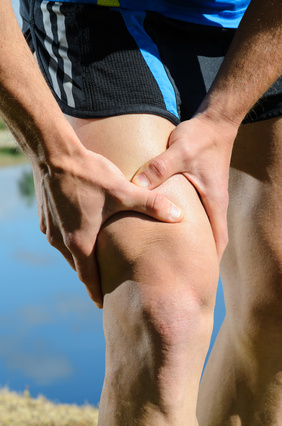
 Knee pain is an extremely common complaint that we treat as physiotherapists and often I get asked "what exactly is wrong with my knee and why is it hurting?"
So I thought that it would be good to do a couple of blog posts about the knee, its anatomy what can become injured, the underlying reasons for some injuries and also how to rehabilitate and strengthen the knee.
Firstly in this blog we will have a look at the underlying anatomy of the knee. Be warned that despite being a simple hinge joint there is a surprisingly large amount of anatomy.
As with all parts of the body, if you want to know what is hurting then you need a good understanding of the anatomy, so we shall now have a look at the anatomy of the knee. The knee is best thought of as being 2 joints - firstly the hinge joint between the femur (upper leg bone) and the tibia (shin bone) and secondly the patella-femoral joint between the patella (kneecap) and femur. Inside of the actual knee joint, the tibial plateau and end of the femur are covered with articular hyaline cartilage which serves to reduce friction and aid with shock absorption.
This is a fairly standard arrangement in all joints in the human body but the knee is special as it has two very special bits of fibro-cartilage called the menisci. The two menisci (lateral and medial) serve to help with shock absorption and improve the congruency (fit) of the joint and are critical parts of a good functioning knee. They are semi-circular in nature and the medial meniscus blends in part with the medial collateral ligament. Inside of the knee joint and in between the femur and tibia are two special and highly important ligaments called the cruciate ligaments.
 The anterior cruciate ligament (ACL) stops the tibia shearing forward away from the femur and the posterior cruciate ligament (PCL) does the opposite. On either side of the knee joint and blending with the capsule of the joint are the collateral ligaments, the medial collateral ligament runs from the femur down to the tibia and provides end of range stability for the knee and the lateral collateral ligament runs from the femur to the fibula. There are also several bursae around the knee that are important to be aware of, a bursa is a gel filled sac that reduces friction and is often found wherever one part of the body such as a tendon runs over another. The pre-patella bursa sits over the top of the patella itself, the infrapatella bursa sits underneath the patella and reduces friction on the patella tendon and finally the suprapatella bursa sits above the patella and underneath the quads tendon.
Finally there is the patella (kneecap) itself which is the largest sesamoid bone in the human body and sits inside of the quadriceps tendon. The patella has articular hyaline cartilage on its under surface to reduce friction and is connected to the tibia by the patella tendon which inserts into the tibial tuberosity on the front of the shin.
There are also a few sets of muscle groups that it is vital to know - the hamstrings and the quadriceps. The hamstrings are a group of three muscles at the back of the thigh - semitendinosus, semimembranosus and biceps femoris, that are responsible for bending the knee backwards and also extending the hip. They run from the ischial tuberosity (this is the bone in the bum that you sit on) to the tibia. The quadriceps are a group of 4 muscles at the front of the thigh - vastus lateralis, vastus medialis, rectus femoris and vastus intermedius, that are responsible for straightening the knee and also joint together to form the quadriceps tendon which joins to the patella.
 Now, despite having covered a fair amount of anatomy, I have actually omitted quite a bit of the anatomy of the knee (I told you that it was complicated!) but I think that I have covered the key points.
Certainly as a physiotherapist seeing patients I would say that the anatomical parts covered above are the key things to understand and be aware of in clinical practice. There are many more bursae situated around the knee and quite a few more muscles but the majority of injuries to the knee happen to the structures described above. Next time we shall have a look at how they get injured and some of the underlying biomechanical issues regarding these injuries.
If you or someone you know currently has knee pain and would like physiotherapy to help resolve it then please get in touch, we can be contacted on 0788 428 1623 or via mail enquiries@threespiresphysiotherapy.co.uk. We provide a home visit physiotherapy service anywhere within 25 minutes drive of Lichfield including areas such as Tamworth, Cannock, Sutton Coldfield, Four Oaks, Burton, Rugeley and Burntwood.
REQUEST A CALLBACK
Just fill in the form below and give us a quick idea of your problem/request so that we can be better prepared to help you.
